 WhatsApp Us (24/7)
 +91 926 888 0303 (24/7)


Abstract: Demyelinating diseases of the brain represent a complex group of neurological disorders characterised by the destruction of the myelin sheath surrounding nerve fibres, which disrupts neural transmission and leads to a wide range of neurological deficits1 . This article reviews the pathophysiology, radiologic features, and diagnostic criteria of key demyelinating disorders, including Multiple sclerosis (MS), Neuromyelitis optica, Myelin oligodendrocyte glycoprotein antibody-associated disease (MOGAD), Tumefactive demyelinating lesions, Acute necrotizing encephalopathy, and Krabbe disease. Advanced imaging techniques, particularly Magnetic resonance imaging (MRI), play a crucial role in the identification and differentiation of these diseases. Key MRI findings, such as T2-weighted hyperintense lesions in specific brain regions, contrast enhancement patterns, and lesion evolution, aid in diagnosis and disease monitoring. Radiologists play a vital role in early diagnosis, monitoring treatment response, and detecting complications, thereby guiding clinical management and improving patient outcomes in demyelinating brain diseases.
Key words: Magnetic Resonance Imaging (MRI), Demyelination, Multiple Sclerosis (MS), Posterior Reversible Encephalopathy Syndrome (PRES), Neuromyelitis Optica, Acute Disseminated Encephalomyelitis (ADEM), Progressive Multifocal Leukoencephalopathy.
Introduction
Demyelinating disorders are a complex group of conditions that disrupt the myelin sheaths of the central nervous system (CNS), affecting white matter and leading to various neurological symptoms1 . These disorders can be classified into several categories based on their causes:
Key categories of demyelinating disorders
- Autoimmune demyelination
- Multiple sclerosis (MS): A chronic autoimmune condition marked by recurrent neurological episodes due to myelin damage2. Subtypes include:
- - Marburg variant: Aggressive with rapid progression
- - Schilder type: Characterised by large, confluent lesions
- - Baló concentric sclerosis: Notable for concentric rings of demyelination
- - Tumefactive demyelinating lesion: Large lesions that can mimic tumours on imaging
- - Neuromyelitis optica spectrum disorder (NMOSD): Primarily impacts the optic nerves and spinal cord
- - Acute disseminated encephalomyelitis (ADEM): An inflammatory demyelinating condition that often follows viral infections
- - MOG-associated disease (MOGAD): Associated with antibodies against myelin oligodendrocyte glycoprotein
- - Acute haemorrhagic leukoencephalitis (Weston Hurst Syndrome): A severe, acute demyelinating process typically following an infection
- Viral demyelination
- Progressive multifocal leukoencephalopathy (PML): Caused by the JC virus, primarily affecting immunocompromised individuals
- Human immunodeficiency virus (HIV)-related white matter changes: - Can include HIV encephalitis, leading to cognitive decline and motor deficits
- Subacute sclerosing panencephalitis (SSPE): A progressive neurological disorder that occurs following measles infection
- Toxic or metabolic demyelination
- Osmotic demyelination: Often occurs due to rapid correction of hyponatremia
- Toxic leukoencephalopathy:s: Associated with substance abuse, particularly inhalation of heroin ("chasing the dragon")
- Mechanical demyelination
- Trigeminal neuralgia: Primarily a pain syndrome, it may involve demyelination of the trigeminal nerve
Understanding these categories helps in the diagnosis and management of demyelinating disorders, as each subtype has distinct clinical features and treatment approaches.
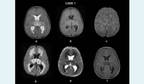
Case 1: A 2-year-old male child presented with altered sensorium with a history of a viral prodrome 8 days back. MRI brain revealed symmetrical areas of T2 (a) and FLAIR (b) hyperintensity involving bilateral thalami as well as periventricular white matter. Gradient echo sequence (GRE) (c) revealed multiple tiny foci of blooming in these areas representing microhaemorrhages. Extensive diffusion restriction was seen in these areas on Diffusionweighted imaging (DWI) (d) with corresponding hypointensity on Apparent diffusion coefficient (ADC) maps (e). No significant contrast enhancement was noted (f). In the given clinical context and the aforementioned MRI findings, possibility of acute necrotizing encephalopathy of childhood (ANEC) was kept and the patient was managed accordingly
Discussion
Acute necrotizing encephalopathy (ANE): ANE is often referred to as acute necrotizing encephalopathy of childhood (ANEC) and is a rare encephalopathy characterised by multiple bilateral brain lesions, primarily affecting the thalami, but also involving the putamina, internal and external capsules, cerebellar white matter, and brainstem tegmentum.3
Pathology:
The precise aetiology and pathogenesis of ANE remain only partially understood. It often follows viral infections, including:
- Influenza A and B
- Parainfluenza
- Varicella
- Enterovirus
- Severe acute respiratory syndrome coronavirus 2 (SARSCoV-2)
MRI findings: Typically shows bilateral symmetrical thalamic involvement with hypointense lesions on T1-weighted images and hyperintense lesions on T2-weighted images. Restricted diffusion is common, and additional findings may include haemorrhage, cavitation, and post-contrast enhancement. MR spectroscopy can indicate elevated lipid/lactate and glutamine/ glutamate peaks.4
Case 2: A 14-year-old male child presented with altered consciousness for the past 2 days. The patient also had a history of viral prodrome 2 weeks back. MRI brain was done which revealed ill-defined patchy areas of asymmetrical T2 and FLAIR hyperintense signal intensity (a and b) involving bilateral thalami as well as right globus pallidus. No abnormal diffusion restriction (c) or susceptibility changes (d) were seen in these lesions. In addition, FLAIR image reveals involvement of the body of corpus callosum (e) as well as right middle cerebellar peduncle & adjoining cerebellar white matter (f). T2 weighted sagittal images of cervical spine also shows faint patchy involvement of the cervical cord. Possibility of a demyelinating etiology was kept. On further evaluation, patient was found to be positive for IgG antibodies against myelin oligodendrocyte glycoprotein (MOG) suggesting diagnosis of MOGAD.
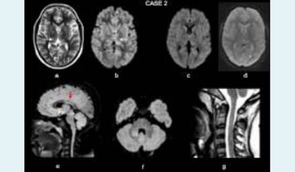
MOGAD is an inflammatory disorder that affects the CNS. It is marked by the presence of IgG antibodies against MOG, a protein found on myelin (the insulating layer around nerves). MOGAD is considered separate from similar conditions like MS, NMOSD, and ADEM, though it shares some features with them.5
Symptoms
- Optic neuritis (41-63%): This is the most common symptom and can cause repeated inflammation of the optic nerve
- Transverse myelitis (30%): Involves inflammation of the spinal cord, sometimes extending across long segments
- ADEM-like encephalomyelitis (2-6%): Causes brain and spinal cord inflammation
- Cortical encephalitis: May cause seizures and is linked with specific brain changes on MRI
- Infratentorial syndromes: Involves the brainstem or cerebellum
Pathology
MOG is found on cells called oligodendrocytes and on the outer layers of myelin sheaths, which protect nerves.
MRI findings: There are no unique MRI signs of MOGAD, but certain features are common.
Brain involvement: Lesions are often few but large, usually in both brain hemispheres, with ill-defined edges. Grey matter and brainstem involvement are more common in children.6
Optic nerve involvement: Over 80% of patients have optic nerve inflammation, often in both eyes, causing nerve swelling and tortuosity. It may also affect the optic sheath and sometimes causes swelling around the eye.
Spinal cord involvement: MOGAD often affects the center of the spinal cord, impacting both grey and white matter. The conus medullaris (lower part of the spinal cord) is commonly involved.
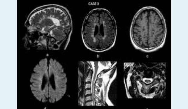
Case 3: A 44-year-old female patient presented with a past history of multiple episodes of sensory and motor neurological deficits. Sagittal T2Wt image (a) shows multifocal T2 hyperintense lesions oriented perpendicular to the ventricular margin (Dawson’s finger). Axial FLAIR image (b) shows multifocal hyperintense lesions oriented perpendicular to the bilateral lateral ventricles in perivenular distribution. Majority of these lesions appear hypointense on T1Wt image (c) likely representing burnt-out lesions. No significant diffusion restriction is seen in the aforementioned lesions as seen on axial DWI image (d). In addition, patchy areas of T2 hyperintense signal intensity is seen involving the cervical cord primarily at C2-C3 vertebral level (e). Axial T2Wt image of cervical cord shows focal area of T2 hyperintense signal intensity along the left posterolateral aspect. Findings are typical of MS.
Multiple sclerosis: It is a relatively common, acquired chronic demyelinating disease affecting the CNS. It is the second most common cause of neurological impairment in young adults, following trauma.7
Key characteristics:
Dissemination: MS is defined by the presence of multiple lesions occurring in different regions of the brain ("disseminated in space") and at different times ("disseminated in time")
Clinical variants: Several clinical variants of MS are recognised, each with distinct imaging findings and clinical presentations:
- Classic multiple sclerosis (Charcot type): This is the most recognised form, characterised by typical relapsingremitting episodes.
- Paediatric-onset multiple sclerosis: Defined variably as MS with onset before the age of 16 to 18 years. It may present differently than adult-onset MS.
- Tumefactive multiple sclerosis: Characterised by large lesions that can mimic tumours on imaging, often causing significant mass effect.
- Marburg type (Acute malignant MS): A rapidly progressive form that leads to significant disability in a short period.
- Schilder type (Diffuse cerebral sclerosis): Involves more widespread lesions and may present with significant cognitive impairment and neurological deficits.
These variants highlight the heterogeneity of MS and the importance of tailored approaches to diagnosis and management.
Epidemiology of MS
- Onset age: The average age of onset is around 29 years, with cases recognized from adolescence into the 60s
- Gender distribution: There is a strong female preponderance
Presentation
-
- Symptoms: The clinical presentation of MS varies based on the location of plaques and may include:
- Sensory or motor disturbances
- Cranial nerve involvement, such as optic neuritis and trigeminal neuralgia
Aetiology of MS
The exact aetiology of MS remains poorly understood, though it is believed to involve both genetic and acquired factors. Several theories and hypotheses regarding its causes include:8
- Infectious agents: There is a longstanding suspicion of an infectious agent, such as Epstein-Barr virus (EBV), due to geographic distribution patterns and clusters of cases. However, no specific infectious agent has been conclusively confirmed as a causative factor.
- Neurotropic viral hypothesis: The presence of symmetric high T2/FLAIR signal changes in the intrapontine segments of the trigeminal nerve in 3-7% of MS patients has raised speculation about a potential neurotropic viral cause stemming from the aerodigestive tract.
- Chronic cerebrospinal venous insufficiency (CCSVI): Some authors have suggested that CCSVI may cause or exacerbate MS, but this theory lacks conclusive evidence from further investigations.
- Autoimmune mechanism: MS is largely considered to result from a cell-mediated autoimmune response targeting the body’s own myelin components. This leads to the loss of oligodendrocytes, with minimal axonal degeneration during the acute phase. However, later stages show significant axonal degeneration following oligodendrocyte loss.
Pathological stages of MS lesions
Demyelination in MS occurs in discrete areas called plaques, which can vary in size from a few millimetres to several centimetres. Each plaque goes through three pathological stages:
- Early acute stage (active plaques):
- Characterised by active myelin breakdown
- Plaques appear pink and swollen due to inflammation
- Subacute stage:
- Plaques become paler ("chalky") as inflammation subsides
- Abundant macrophages are present, indicating ongoing damage
- Chronic stage (inactive plaques/Gliosis):
- Minimal to no myelin breakdown occurs
- Gliosis leads to volume loss in the affected areas, resulting in plaques that appear grey or translucent
Classification
Clinically isolated syndrome (CIS): An isolated neurological deficit with a solitary plaque on imaging; progression to MS is uncertain.
- Relapsing-Remitting MS (RRMS): Accounts for about 70% of cases, characterised by periods of symptoms (relapses) followed by complete resolution.
- Secondary Progressive MS (SPMS): Progression of symptoms occurs after an initial relapsing-remitting phase, with a decline in baseline function.
- Primary Progressive MS (PPMS): Progressive deterioration from the time of diagnosis, typically with little to no remission
Pathophysiology
- Demyelination: MS involves white matter demyelination visible as plaques on MRI, with damage primarily to the myelin sheath surrounding axons and minimal underlying neuronal damage
- Autoimmune component: The condition is likely autoimmune and cellular-mediated, although no unifying cause or trigger has been identified. Some genetic and environmental factors may contribute
Investigation
- MRI: MRI is the investigation of choice for MS; it should be performed with contrast and include spinal imaging to evaluate the extent of disease involvement and monitor plaque progression
MRI in MS: MRI has transformed the diagnosis and monitoring of MS. It not only aids in confirming the diagnosis (as per the McDonald diagnostic criteria) but also helps evaluate treatment response and determine disease patterns through follow-up scans.9, 10
Plaque locations: Plaques can occur in various locations, including:
- Infratentorial
- Deep white matter
- Periventricular
- Juxtacortical
- Mixed white matter-grey matter lesions
Predictive features
Even a single MRI scan can help differentiate between relapsingremitting and progressive disease forms. Features favouring progressive disease include:
- Large, numerous plaques
- Hyperintense T1 lesions
These imaging characteristics play a crucial role in the management and treatment planning for individuals with MS.
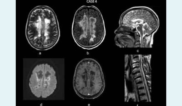
Case 4: A 41-year-old female presented with gradually progressive weakness in bilateral upper and lower limbs. Contrast enhanced MRI of the brain reveals confluent areas of T2 (a) and FLAIR (b) hyperintense signal intensity in bilateral periventricular white matter. There is partial central signal suppression on FLAIR. Sagittal T2Wt image (c) shows diffuse involvement of the corpus callosum. Axial diffusion weighted image (d) reveals mild peripheral diffusion restriction on both sides. No significant contrast enhancement is seen on axial T1Wt post-contrast image (e). T2Wt sagittal image of the cervical spine reveals longitudinally extensive T2 hyperintense signal intensity involving the cervical cord. Mildly reduced bulk of the cervical cord is also noted. Findings are likely suggestive of a chronic demyelinating etiology. Further workup revealed antiAQP4-IgG positivity suggesting Neuromyelitis optica.
Discussion
Neuromyelitis Optica Spectrum Disorder (NMOSD): It is a severe demyelinating disease primarily characterized by the presence of autoantibodies targeting the aquaporin-4 (AQP4) water channel in seropositive cases.11
Classic presentation
The classic clinical triad for NMOSD includes:
- Bilateral optic neuritis: Inflammation of the optic nerves, often resulting in vision loss.
- Longitudinally extensive transverse myelitis: Severe inflammation affecting a long segment of the spinal cord, leading to neurological deficits.
- Positive anti-AQP4 antibody test: Detection of antibodies against AQP4, confirming the diagnosis in seropositive individuals.
Expanded spectrum of manifestations
While the classic triad is significant, NMOSD encompasses a broader range of symptoms and manifestations, highlighting the complexity and variability of the disorder. These may include:
- Other forms of optic neuritis
- Brainstem syndromes
- Area postrema syndrome (nausea and vomiting)
- Cortical or subcortical lesions
Understanding the diverse clinical presentations of NMOSD is crucial for accurate diagnosis and effective management.
Early disease characteristics
In the early stages of NMOSD, the pathological findings resemble those seen in multiple sclerosis, including:
- Macrophage and microglia activation: Indicative of an inflammatory response
- Axonal damage: Damage to axons occurs early and is notable as it precedes demyelination in NMOSD
Pathology of NMOSD
In approximately 70% of patients with established NMOSD, antiAQP4-IgG antibodies can be detected. These antibodies target the aquaporin-4 (AQP4) water channel, which is predominantly located on astrocyte foot processes, particularly around the circumventricular organs, such as the periaqueductal gray matter.12
Aetiology
While NMOSD is generally sporadic, there is some evidence suggesting immunogenic similarities between certain viral infections and the aquaporin-4 water channel, potentially triggering the autoimmune response in susceptible individuals. Understanding these pathological and immunological features is crucial for diagnosing and treating NMOSD effectively.13
MRI in NMOSD
MRI Overview
MRI is the primary imaging modality for diagnosing NMOSD, and both the orbits, brain, and spinal cord should be examined in suspected cases.14
Orbits
- Optic Neuritis Features:
Optic nerves appear hyperintense and swollen on T2- weighted sequences
- Enhancement on T1-weighted images with contrast (T1 C+)
- Bilateral optic nerve involvement extending to the chiasm is particularly indicative of NMOSD
- Chronic stages may show atrophy and hyperintensities
Brain
- Lesion characteristics:
Aquaporin-4 distribution: Lesions are typically found in areas rich in aquaporin-4, such as:
- Periventricular regions
- Periaqueductal gray matter
- Hypothalamus and medial thalamus
- Dorsal pons and medulla
- Corpus callosum with a marbled appearance.
- Longitudinal lesions: Involvement of the corticospinal tract and larger hemispheric white matter lesions (>3 cm)
- Cystic changes: Some lesions may resolve, but cystic changes can occur in a minority of cases
Chronic changes:
- Longstanding disease may show extensive bilateral symmetric T2/FLAIR hyperintensities involving subcortical and deep white matter
Spinal cord
- Longitudinally extensive lesions:
- High T2 signals spanning at least three vertebral segments, indicating extensive spinal cord involvement
- Swelling is usually present in acute phases
- Imaging features:
- T1: Hypointense with potential for cord atrophy in followup scans
- T2: Hyperintense lesions, often >3 vertebral body lengths, with specific bright spotty lesions
- T1 C+: Variable enhancement patterns, including ringenhancement and patchy "cloud-like" appearances
Distinguishing features of NMOSD from MS
While no features are exclusively diagnostic, several imaging characteristics can help differentiate NMOSD from MS. These distinctions may be particularly relevant in Asian populations, where overlap can be more common.
Brain imaging differences
- Lesion distribution:
- NMOSD exhibits smooth, confluent periependymal lesions
- MS typically shows oval lesions oriented perivenularly (Dawson fingers)
- Juxtacortical lesions:
- Fewer juxtacortical (U-fiber) lesions in NMOSD
- Corpus callosum:
- NMOSD may show more extensive involvement of the corpus callosum, especially its ependymal surface
- Lesion size and shape:
- NMOSD lesions are often larger and more confluent
- Enhancement patterns:
- NMOSD lacks open ring enhancement, which is sometimes seen in MS
- Corticospinal tract and diencephalon:
- More frequent involvement of these areas in NMOSD
Spinal cord imaging differences
- Lesion extent:
- NMOSD typically presents with more longitudinally extensive spinal cord lesions
- Cord involvement:
- NMOSD favours central spinal cord involvement over peripheral regions
Optic Nerve Differences
- Optic Neuritis:
- NMOSD features more longitudinally extensive optic neuritis with a preference for the posterior optic pathway.
These imaging characteristics can aid in differentiating NMOSD from MS, guiding appropriate diagnosis and treatment.
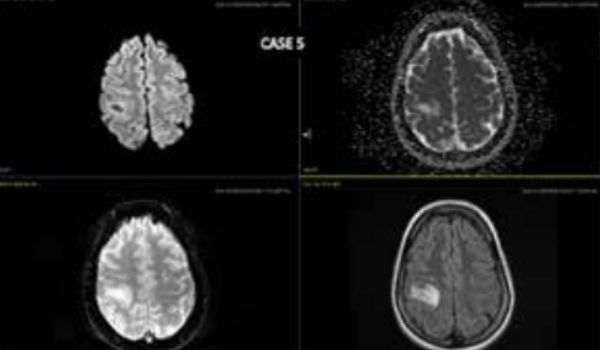
Case 5: A 60-year-old male presented with seizure and difficulty in speech. Contrast enhanced MRI of the brain demonstrates hyperintensity on T2W and FLAIR, with mild restricted diffusion and no blooming on GRE. There is no mass effect seen as expected for the lesion size
On MRS imaging, it reveals mildly elevated peak of choline. On T1 Gd+, the lesion shows faint open ring enhancement. Findings (Cho) are likely suggestive of tumefactive demyelinating lesion.
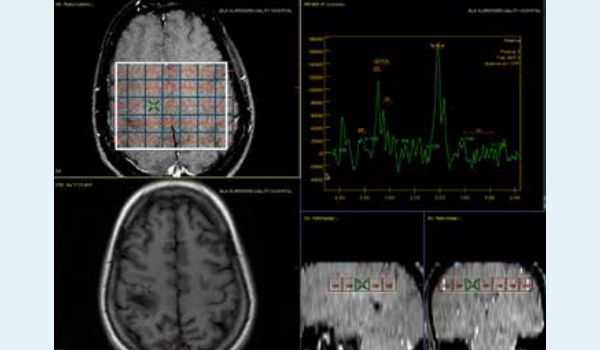
Magnetic resonance spectroscopy (MRS) imaging
Discussion
Tumefactive demyelination
Definition: Tumefactive demyelinating lesions (TDLs), also known as monofocal acute inflammatory demyelination (MAID), are aggressive forms of demyelination that typically present as solitary lesions (or occasionally a few) larger than 2cm, often mimicking tumours on imaging.15
Imaging characteristics:
- Mass effect:
TDLs usually exhibit minimal mass effect and surrounding oedema
- Contrast enhancement:
They typically show an open-ring pattern of enhancement after contrast administration
- Diffusion characteristics:
High apparent diffusion coefficient (ADC) values are common
- Cerebral blood volume:
Low relative cerebral blood volume (rCBV) is typically observed
These features can help differentiate TDLs from neoplasms and guide appropriate clinical management.
MRI features of TDLs
Classification: The International Magnetic Resonance Imaging in MS Collaboration categorizes TDLs into several appearances:
- Ring-enhancing
- Diffusely infiltrative
- Megacystic
- Baló-like
General characteristics:
- TDLs are typically large with relatively little mass effect or surrounding edema
- Centrally located dilated veins may be observed within these lesions
Contrast imaging (T1 C+):
- Approximately 50% of TDLs show contrast enhancement
- The enhancement pattern typically appears as an open ring, with the incomplete portion located on the grey matter side of the lesion
Perfusion imaging:
- Helps differentiate TDLs from high-grade gliomas and lymphomas
- TDLs demonstrate significantly lower mean relative cerebral blood volume compared to these tumours
Diffusion imaging (DWI):
- Less effective in differentiating TDLs from necrotic tumours, as both may show increased apparent diffusion coefficient (ADC)
- However, DWI can help distinguish ring-enhancing TDLs from cerebral abscesses, as abscesses show restricted diffusion while TDLs show mildly increased ADC.
MR Spectroscopy:
- Specific to TDLs is the presence of elevated beta, gamma glutamate/glutamine (β,γ-Glx)
- While increased choline and lactate may support the diagnosis of TDLs, they are not specific
- The metabolic pattern may resemble that of neoplasms, with decreased NAA/Cr ratio and increased Cho/Cr ratio, along with variable lactate and lipid peaks
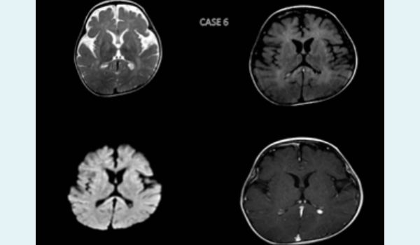
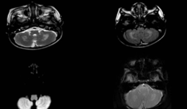
Case 6: A 6-month-old female child presented with irritability, fever, and hypertonia. Contrast-enhanced MRI of the brain symmetric T2/FLAIR hyperintensities in bilateral periventricular and juxta-cortical regions, posterior limb of bilateral internal capsules, crus cerebri and along bilateral cortico-spinal tracts.
T2/FLAIR hyperintensities are also seen in bilateral cerebellar hemispheres, dentate nuclei.
Discussion
Myelin-producing cells degenerate in Krabbe disease, also known as globoid cell leukodystrophy, an autosomal recessive lysosomal storage illness. Because myelin turnover is disrupted, this illness affects both the central and peripheral nerve systems, resulting in a range of neurological symptoms.16 The condition is characterized by the buildup of toxic chemicals in cells, which specifically affects the Schwann cells and oligodendrocytes that make myelin.17
Features of Krabbe Disease on Radiographs:
CT Findings:
Early alterations: At first, CT may reveal symmetrically hyperdense regions including:
- Thalami and cerebellum
- Caudate nuclei
- Internal capsule's posterior limbs
- The brainstem
- Potential expansion into the semiovale centrum/corona radiata
Subsequent alterations: Hypoattenuation occurs in the semiovale's white matter.
- The cerebellum and cerebrum gradually atrophy
MRI findings
- T2 Imaging
- High signal intensity is seen predominantly in the parietooccipital regions or diffusely in periventricular white matter
- Involvement of corticospinal tracts, including internal capsules and brainstem
- Subcortical U-fibers may remain intact until late stages
- A "tigroid" pattern of white matter involvement may be observed
- T1 C+ (Gd): Typically shows no contrast enhancement in affected areas
- MR Spectroscopy (MRS):
- The adult form may demonstrate abnormal choline elevation in the centrum semiovale
- Additional findings:
- Enhancement and enlargement of the optic nerves and, occasionally, peripheral nerve
Conclusion:
ANE and the other demyelinating disorders discussed—MOGAD, MS, NMOSD, tumefactive demyelination, and Krabbe disease— highlight the complexity of neurological pathology. ANE, typically following viral infections, is characterised by severe bilateral lesions primarily in the thalami,3 while MOGAD presents with inflammatory symptoms affecting the optic nerves and spinal cord.5 MS is notable for its heterogeneity and varying clinical presentations, driven by an autoimmune mechanism that leads to demyelination.
NMOSD, distinguished by the presence of anti-AQP4 antibodies, showcases a different pathophysiological mechanism, leading to unique MRI findings that assist in its diagnosis.11 Tumefactive demyelinating lesions challenge clinical diagnosis due to their tumour-like appearance, necessitating advanced imaging techniques for differentiation. Lastly, Krabbe disease illustrates the impact of genetic disorders on myelin production, resulting in widespread neurological impairment.
Collectively, these conditions underscore the importance of precise imaging and clinical evaluation in diagnosing and managing demyelinating and encephalopathic disorders. Understanding their distinctive features not only aids in accurate diagnosis but also informs treatment strategies, improving patient outcomes in this diverse spectrum of neurological diseases
Understanding the unique characteristics, imaging findings, and clinical implications of these conditions is essential for accurate diagnosis, timely intervention, and effective management, ultimately improving patient outcomes. Continued research into their aetiologies and treatment options remains vital in the ever-evolving field of neurology.
Shashank Raj, Prem Kumar Ganesan, Vivek Kasana, Densia Rons Thampi. MRI Findings in Demyelinating
Diseases of the Central Nervous System. MMJ. 2024, Dec. Vol 1 (4).
DOI: XXXXX
References
- Love S. Demyelinating diseases. Journal of clinical pathology. 2006;59(11):1151-9.
- Sarbu N, Shih RY, Jones RV, et al. White matter diseases with radiologic-pathologic correlation. Radiographics. 2016;36(5):1426-47.
- Mizuguchi M, Abe J, Mikkaichi K, et al. Acute necrotising encephalopathy of childhood: a new syndrome presenting with multifocal, symmetric brain lesions. Journal of Neurology, Neurosurgery & Psychiatry. 1995;58(5):555-61.
- Wang HS, Huang SC. Acute necrotizing encephalopathy of childhood. Chang Gung medical journal. 2001;24(1):1-0.
- Reindl M, Rostasy K. MOG antibody-associated diseases. Neuroimmunology & Neuroinflammation. 2015;2(1):e60.
- Dos Passos GR, Oliveira LM, Da Costa BK, et al. MOG-IgGassociated optic neuritis, encephalitis, and myelitis: lessons learned from neuromyelitis optica spectrum disorder. Frontiers in neurology. 2018;9:217.
- Gupta S, Tewari A, Nair V. Multiple sclerosis: Indian perspective. API India Medicine Update. 2013:547-51.
- Thompson AJ, Banwell BL, Barkhof F, et al. Diagnosis of multiple sclerosis: 2017 revisions of the McDonald criteria. The Lancet Neurology. 2018;17(2):162-73.
- Simon JH, Li D, Traboulsee A, et al. Standardized MR imaging protocol for multiple sclerosis: Consortium of MS Centers consensus guidelines. American Journal of Neuroradiology. 2006;27(2):455-61.
- Lövblad KO, Anzalone N, Dörfler A, et al. MR imaging in multiple sclerosis: review and recommendations for current practice. American journal of neuroradiology. 2010;31(6):983-9.
- Wingerchuk DM, Lennon VA, Lucchinetti CF, et al. The spectrum of neuromyelitis optica. The Lancet Neurology. 2007;6(9):805-15.
- Kim SH, Kim W, Li XF, et al. Clinical spectrum of CNS aquaporin-4 autoimmunity. Neurology. 2012;78(15):1179-85.
- Huh SY, Min JH, Kim W, et al. The usefulness of brain MRI at onset in the differentiation of multiple sclerosis and seropositive neuromyelitis optica spectrum disorders. Multiple Sclerosis Journal. 2014;20(6):695-704.
- Bennett JL. Finding NMO: the evolving diagnostic criteria of neuromyelitis optica. Journal of Neuro-Ophthalmology. 2016;36(3):238-45.
- Algahtani H, Shirah B, Alassiri A. Tumefactive demyelinating lesions: a comprehensive review. Multiple sclerosis and related disorders. 2017;14:72-9.
- Farina L, Bizzi A, Finocchiaro G, et al. MR imaging and proton MR spectroscopy in adult Krabbe disease. American Journal of Neuroradiology. 2000;21(8):1478-82.
- Wenger DA, Luzi P. Krabbe disease: globoid cell leukodystrophy. InRosenberg's molecular and genetic basis of neurological and psychiatric disease. Academic Press. 2025;593-603.