 WhatsApp Us (24/7)
 +91 926 888 0303 (24/7)


Abstract:
Epiphrenic diverticulum is a rare type of pulsion diverticulum that develops in the lower third of the oesophagus due to increased intra-oesophageal pressure. Patients often present with symptoms such as dysphagia, retrosternal discomfort, and recurrent vomiting or regurgitation of food. Surgery is considered the most effective treatment that addresses both the high intra-oesophageal pressure and the diverticulum itself. Traditionally, this procedure was performed via thoracotomy, which involved significant morbidity. However, recent advances in minimally invasive techniques, have led to a shift from a thoracoscopic to a laparoscopic approach for treating majority of these diverticula. We present a case of a 75-year-old male who was initially misdiagnosed with a sliding hiatus hernia. He underwent hiatus repair and fundoplication, but his symptoms persisted, prompting further evaluation, which revealed an epiphrenic diverticulum. The previous surgical intervention was reversed, and a laparoscopic excision of the epiphrenic diverticulum with cardiomyotomy and anterior fundoplication was done. The patient recovered well postoperatively and was able to eat without any difficulty
Key words: Epiphrenic Diverticulum, Laparoscopic Cardiomyotomy
Introduction
Oesophageal diverticulum is a rare condition, classified into two types – pulsion and traction diverticula. Pulsion diverticula are false diverticula (containing only mucosa and submucosa) while traction diverticula are true diverticula, containing all layers of the oesophageal wall. Pulsion diverticulum, as the name suggests, arise due to increased intra-oesophageal pressure, often secondary to an obstruction distal to the diverticulum. Diverticula at the upper end of the oesophagus are due to increased pressure in the cricopharyngeal sphincter and are known as Zenker’s diverticula. Ephiphrenic diverticula, located at the lower end of the oesophagus result from increased pressure in the lower oesophageal sphincter (LES), resulting in an outpouching of the mucosa and submucosa through the muscular layer of the oesophagus.1
The majority of patients are symptomatic primarily due to obstructive features than the diverticulum itself, presenting with dysphagia, retrosternal discomfort and regurgitation or vomiting.2-4 Surgery is the preferred treatment method, addressing both increased oesophageal pressure and the diverticulum. Minimally invasive surgery is the method of choice, with the laparoscopic intra-abdominal approach increasingly favoured3-5 as it effectively addresses both components of the surgery. Additionally, fundoplication can be performed to prevent reflux following cardiomyotomy. Morbidity and mortality rates have significantly reduced after adoption of the intraabdominal laparoscopic approach. However, in certain cases, a thoracoscopic approach may still be required to excise the diverticulum.
Case report
A 75-year-old male presented with a sensation of retrosternal sticking of food, especially with solids, for the last few months. This sensation had progressively worsened, limiting him to eat a semisolid food. He was evaluated externally, where a contrastenhanced computed tomography (CECT) scan of the chest and abdomen was done. The CT report suggested a sliding hiatus hernia with herniation of stomach into the thorax. As the patient was apprehensive about undergoing an endoscopy, he was operated, based solely on the CT findings, with laparoscopic hiatus hernia repair and fundoplication.
Postoperatively, his symptoms worsened to the point where he could only tolerate a liquid diet. He presented to us with these complaints and was advised to undergo an upper gastrointestinal (GI) endoscopy. The endoscopy revealed a large diverticulum at the lower end of the oesophagus (Figure 1). The CT was subsequently reviewed at Max hospital, revealing that what had been reported as herniation of the stomach above the diaphragm was, in fact, an oesophageal diverticulum arising from the left wall of the oesophagus just above the diaphragm (Figure 2).
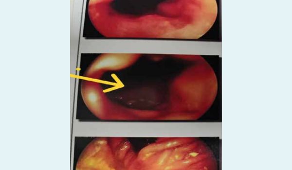
Figure 1: Endoscopy showing diverticulum mouth marked with arrow.
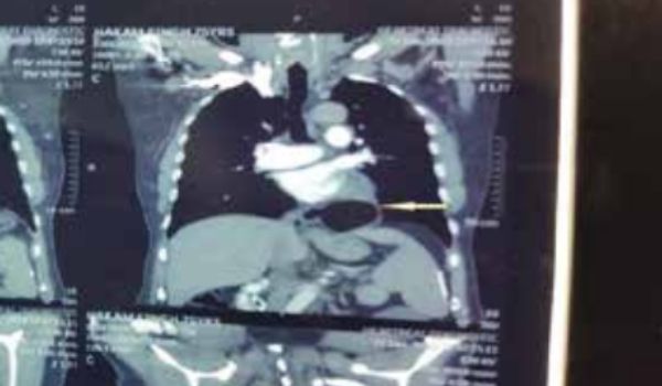
Figure 2a: CECT sagittal section – diverticulum marked with white arrow.
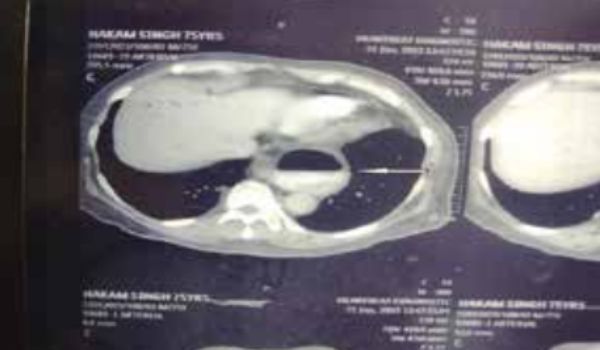
Figure 2b: Previous fundoplication.
After further evaluation, the patient was taken up for surgery again. Laparoscopy revealed the previous wrap of the stomach around the lower end of the oesophagus and evidence of the previous hiatus repair (Figure 3).
Figure 3: Previous fundoplication
Dense adhesions were observed around the hiatus. The stomach wrap was undone, and the lower end of the oesophagus was mobilised into the thorax. The diverticulum was also mobilised with both blunt and sharp dissection and delivered into the abdomen through the diaphragmatic hiatus. An intraoperative endoscopy confirmed the presence of the diverticulum and ruled out any mucosal abnormalities within it. The endoscope was passed beyond the diverticulum into the stomach and the diverticulum was excised using an EndoGIA stapler with the endoscope in situ to prevent narrowing of the oesophagus (Figure 4).
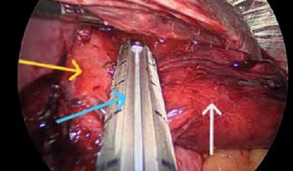
Figure 4: Showing diverticulum ( white arrow) being excised with stapler at neck of diverticulum ( blue arrow) with endoscope in oesophagus ( yellow arrow).
Subsequently, a cardiomyotomy was performed, extending from the base of the diverticulum to 1 - 2cms onto the stomach, to decrease lower oesophageal pressure distal to the diverticulum (Figure 5).
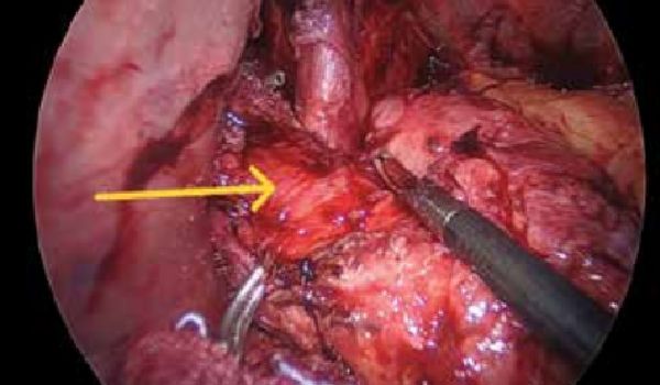
Figure 5: Cardiomyotomy with mucosa pouting out marked with arrow.
The cardiomyotomy was covered with Dor (anterior) fundoplication to prevent reflux, which could occur due to impaired lower oesophageal tone following the cardiomyotomy (Figure 6)
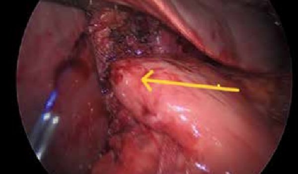
Figure 6: Anterior fundic wrap over cardiomyotomy
Post-operatively, the patient was started on a liquid diet from day 1. A Gastrograffin dye study revealed free flow of dye across the gastro-oesophageal (GE) junction (Figure 7). Now, four months post-surgery, the patient is able to tolerate a normal diet without any difficulty. He complains of occasional reflux, that is manageable with a combination of a proton pump inhibitor and domperidone.
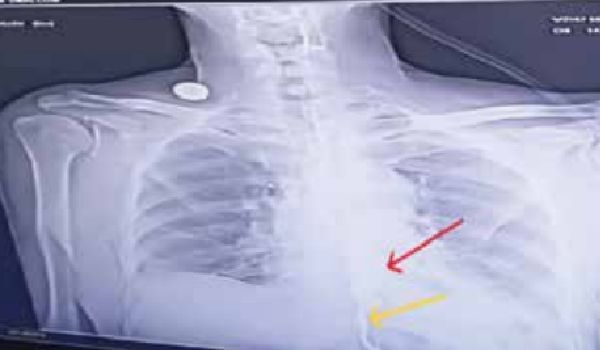
Figure 7: Postoperative – Gastrograffin study showing free flow of contrast across the gastro-oesophageal (GE) junction (yellow arrow) and intact site of excision of diverticulum (red arrow).
Discussion
Ephiphrenic diverticulum is rare type of pulsion diverticulum arising from the distal third of the oesophagus. It is thought to result from increased intraluminal pressure due to hypercontractility and outflow obstruction, with oesophageal dysmotility being the primary cause in most cases.1
The main symptoms include dysphagia, regurgitation, weight loss, heartburn, respiratory complaints, and retrosternal pain when swallowing. Patients are often asymptomatic, but they may suffer from oesophagitis, bleeding from ulceration, impaction, and stasis with regurgitation.2 These are mainly symptoms of the underlying motility disorder rather than the diverticulum itself.3,4
Treatment of diverticulum involves 3 components:
- Correction of the motility disorder, e.g., myotomy
- Excision of the diverticulum
- Fundoplication to address reflux caused by cardiomyotomy
Surgery is also advised in asymptomatic patients due to
Traditionally, the transthoracic approach was used for diverticulum excision. However, recently, multiple series have shown the feasibility of the laparoscopic transhiatal approach with good results.3,4,6,7 The main disadvantage of the laparoscopic approach is the occasional inability in reaching the upper end of the diverticulum transhiatally. Factors that may necessitate a transthoracic approach have been described, allowing for a combined thoracoscopic and laparoscopic approaches for easier diverticulum excision.8 Some series have also described good results with peroral endoscopic myotomy (POEM), an endoscopic procedure that decreases the lower oesophageal sphincter pressure9,10 approach is the occasional inability in reaching the upper end of the diverticulum transhiatally. Factors that may necessitate a transthoracic approach have been described, allowing for a combined thoracoscopic and laparoscopic approaches for easier diverticulum excision.8 Some series have also described good results with peroral endoscopic myotomy (POEM), an endoscopic procedure that decreases the lower oesophageal sphincter pressure9,10
A high index of suspicion and thorough investigation are essential for effective treatment of the oesophageal diverticulum as demonstrated in this case. The laparoscopic approach is feasible and provides satisfactory outcomes with minimum morbidity.
Manmohan Singh Bedi, J. Jose. Epiphrenic Diverticulum Excision by Laparoscopic Transhiatal Approach –
A Case Report. MMJ. 2024, December. Vol 1 (4).
DOI: XXXXXXXX
References
- Nehra D, Lord RV, DeMeester TR, et al. Physiologic basis for the treatment of epiphrenic diverticulum. Ann Surg. 2002;235:346-54.
- Tedesco P, Fisichella PM, Way LW, et al. Cause and treatment of epiphrenic diverticula. Am J Surg. 2005;190:891-4.
- Thomas ML, Anthony AA, Fosh BG, et al. Esophageal diverticula. Br J Surg. 2001;88:629-42.
- Herbella FA, Patti MG. Achalasia and epiphrenic diverticulum. World J Surg. 2015;39:1620-4.
- Yoshida T, Hashimoto S, Mizuno KI, et al. Advanced squamous cell carcinoma in an asymptomatic, large, epiphrenic esophageal diverticulum. Clin J Gastroenterol. 2020;13:477-82.
- Altorki NK, Sunagawa M, Skinner DB. Thoracic esophageal diverticula. Why is operation necessary? J Thorac Cardiovasc Surg. 1993;105:260-4.
- Zaninotto G, Parise P, Salvador R, et al. Laparoscopic repair of epiphrenic diverticulum. Semin Thorac Cardiovasc Surg. 2012;24:218-22.
- Sato Y, Tanaka Y, Ohno S, et al. Optimal surgical approaches for esophageal epiphrenic diverticulum: literature review and our experience. Clinical Journal of Gastroenterology. 2023;16(3):317-24.
- Demeter M, Bánovčin Jr P, Ďuriček M, et al. Peroral endoscopic myotomy in achalasia and large epiphrenic diverticulum. Digestive Endoscopy. 2018;30(2):260-2.
- Yang J, Zeng X, Yuan X, et al. An international study on the use of peroral endoscopic myotomy (POEM) in the management of esophageal diverticula: the first multicenter D-POEM experience. Endoscopy. 2019;51(04):346-9.