

Abstract: The gold standard for cemented hip replacement is the Charnley lowfriction hip replacement. Orthopaedic therapies, such as total hip replacement, have a remarkably high success rate. It relieves patients from pain and improves walking ability and the overall quality of life. We report a 30-year follow-up of our case, a total hip replacement done by a senior joint replacement surgeon at our hospital. A 34-year-old patient, teacher by profession, who was a case of bilateral secondary hip arthritis to Ankylosing Spondylitis, was bed-bound and severely disabled and was operated with total hip replacement in 1995. Long-term follow-up based on functional Harris Hip Score, radiographic status and patient-related outcome measures (PROM). We conclude that Charnley’s cemented total hip replacement has given a time-tested long-term result and is a gold standard surgery if it is done following proper technique.
Key words: Charnley low friction replacement, hip arthritis, ankylosing spondylitis, longevity of THR
Introduction
Total hip replacement is one of the most successful orthopaedic procedures. It relieves patients from pain and improves walking ability and the overall quality of life[1]. As more biological types of treatment for the commonest hip disease, osteoarthritis, are not yet in clinical practice, THA will remain the best treatment available[2]. Only the Charnley LFA with cement has a longer follow-up, with reported series with a minimum 25[3,4] and 30[5,6,7] years results.
The Charnley low-friction replacement is considered the most reliable method for cemented hip replacement[8]. More than 50 years ago, when John Charnley introduced his new method of total hip replacement (THA), he wrote, “Neither surgeons nor engineers will ever make an artificial hip joint which will last 30 years”[8]. The fundamental principles of LFA are the use of a trans-trochanteric approach, a small head of 22.225 mm diameter, a thick all-polyethylene acetabular component, a Monoblock femoral component and cement for fixation[9].
Case: A 34-year-old male patient presented with pain and restriction of movement in both hips and back pain with an antalgic gait. The patient was undiagnosed and was taking treatment from a local practitioner in the form of pain control medication and physiotherapy. X-ray of the pelvis with both hips showed bilateral arthritic hips, bilateral sacroiliitis, and whiskering of bilateral ischial tuberosities. On clinical examination, hip rotations were painful and restricted, the Figure of 4 test was found to be positive, with severe restrictions on spine movements. The patient was initially investigated with an inflammatory arthritis profile which showed HLA B27 positive and the patient was diagnosed as ankylosing spondyloarthropathy. The Senior joint replacement surgeon planned for sequential total hip replacement with Charnley low friction replacement at a span of three months to provide the patient with a functionally mobile and pain-free hip to perform the activities of daily living. After getting the fitness for surgery, the patient was operated on in the lateral position.
Operative procedure
Hip exposed with Harding approach, capsule was cut and hip was dislocated anteriorly. Offset was measured using a scale and a neck cut was planned. The neck cut was done. The acetabulum was exposed and prepared with serial reaming maintaining the native anteversion and inclination angle with reamers. Small drill holes were made in the acetabulum, the copious saline wash was given and the acetabulum was dried, cementing was done in a doughy stage with finger packing, then a cemented 40 mm diameter acetabular component of high molecular weight polyethylene (HMWP) was put in and held with a special instrument until cement was hard. Femur preparation for stem insertion was done with serial straight hollow femoral reamers with increased sizes, maintaining anteversion in the femur. A trial was done followed by an assessment of hip rotation and stability. The loose cancellous bone was removed from the canal, a thorough wash was given and packed with a rolled gauze to dry the canal, a Bone plug was made and put as a cement restrictor in the canal and cementing was done with finger packing following which cemented double tapered monobloc Charnley’s stem was put in anteversion, maintaining the offset of the femur. A metal head of size 22.25 was put in according to the principles of Charnley’s low-friction total hip replacement. Hip was reduced and stability was checked in all movements and was found to be stable. The wound was closed in layers and the procedure was uneventful. The other side was operated on with similar steps after 3 months of the first surgery.
The patient was mobilised for full weight bearing with walker support and physiotherapy was advised for strengthening of the muscles around the hip. The patient’s post-operative x-rays were taken which showed the implant in position and no limb length discrepancy. The patient had to have a good hip range of movement and no pain on subsequent follow-up visits. At three months follow-up, patient was allowed to resume his teaching job. The patient was followed up with a functional Harris Hip score and radiographs, every three months for the first year then 6 monthly the next year and was found to be doing well with a good (80-89 points) Harris Hip Score and excellent patient-related outcome measures (PROMs). The patient then was followed up every 5 years after the surgery and at 30 years with no significant complaint attributing to the hips.
Discussion
The Cemented Total Hip replacement has shown long-term survival results in various literature. We have a case that was operated on by a senior joint replacement surgeon in the year 1995 for bilateral total hip replacement done in a young patient with bilateral secondary degenerative arthritis of the hip with ankylosing spondylitis. On clinical and radiological evaluation done after a 30-year follow-up, the hips are found to be stable with no signs of aseptic loosening or bone resorption present on the X-ray and good Harris Hip Score and patient-related outcome measures (PROMs). The primary hurdle to the longterm survival of total hip replacement is loosening. Loosening was defined as the migration or presence of a continuous clear zone at the bone-cement interface, using the modified criteria of Hodgkinson et al10 for the socket and Harris et al11 for the stem. Thus, the incidence of revision surgeries in the hip has risen in the recent past and this creates the need to study how can we increase the longevity of the total hip replacement by studying the surgical techniques and changes in the implant design. Cemented total hip replacement has been less popular in current practice despite the long-term results that were seen with cemented total hip replacement in the past. The progression of femoral cementing procedures is traditionally characterised in terms of generations[12,13]. Several studies have shown that improved cementing techniques[14] have resulted in a high success rate for joint replacements. One study found an 88% survival rate after 18 years[15], while another study reported an 80% survival rate over 25 years when considering the need for femoral component replacement due to aseptic loosening as the endpoint in second-generation procedures. Williams et al[16] reported a 100% survival rate over a period of 12 years by replacing the femoral component to treat aseptic loosening. This outcome was accomplished through the utilisation of a refined tapered stem and contemporary cementing techniques[17]. The study found that there was a 100% survival rate over a period of 15 years when utilising advanced techniques to replace the femoral component for aseptic loosening. In a study conducted by de Jong and colleagues[18], they found that the survival rate at 20 years was 99% when using revision of the acetabular component as a treatment for aseptic loosening. The procedure involved reaming to the depth of the subchondral layer, creating 6-8 anchorage holes and filling them with cancellous bone using second-generation techniques. Wellcemented components following proper cementing techniques surely decrease the number of revision surgeries. In acetabulum good pre-operative planning (e.g. Osteophytes, need for bone graft, etc) and meticulous acetabular preparation with sequential reamers at least 2 sizes larger than the cup size is required. In the cancellous bone bed, which is created, multiple small anchoring holes have to be made in the roof with a flexible drill. Using pulsatile lavage, the loose cancellous bone has to be removed from the prepared acetabulum and thorough bone bed cleansing is mandatory following which acetabulum is packed with dry gauze to minimise the bleeding and cementing is begun in the doughy stage at higher viscosity to prevent blood lamination at the cement bone interface. Cement is pressurised manually followed by a designed pressuriser. The size of the acetabular component should be at least 2 sizes than the last reamer used to maintain a cement mantel of 2mm, held in the position till curing of the cement is completed. The femoral stem cementing technique also includes preoperative planning to appreciate the femoral anatomy in ap and lateral views followed by planning of the neck cut at 45 degrees to the femoral diaphysis. Entry is identified by the pyriformis fossa and posterolateral canal preparation to prevent posteriorising the tip of the stem. Serial reaming is to be done to prepare the canal for stem insertion followed by thorough pulsatile lavage to remove the loose cancellous bone and preservation of strong cancellus bone. Bone plug inserted 1.5-2 cm distal to the tip of the femur component to create a cement column of 2 cm distally. The canal is washed and dried with roller gauze to prevent the back bleeding from the interface.
Cementing is done with hand packing or gun in a retrograde fashion till the proximal end and the canal opening are then occluded and the cement column is pressurised for 2-3 minutes before stem insertion for proper cement interdigitation in the strong cancellous bone. The planned stem which will maintain the cement mantle of 5 mm medial calcar and 2-3 mm distally is put slowly using a stem introducer which helps to control the version and held in the position till the cement cures. Cemented Total hip replacement continues to be a successful procedure if done following proper techniques. Proper cementing technique holds prime importance rather than the mere choice of implant and if done technically well then long-term survival of the components can be accounted for.
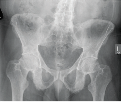
Figure 1: Pre-operative X-rays showing bilateral arthritic hips, bilateral sacroiliitis and ‘whiskering’ of bilateral ischial tuberosities
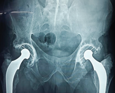
Figure 2: 25-year follow-up X-ray showing operated bilateral cemented total hip replacement with the implant in situ
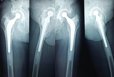
Figure 3: 30-year follow-up X-rays showing operated cemented total hip replacement with the implant in situ. (a) AP view right hip, (b) Lateral view right hip, (c) AP view left hip, (d) Lateral view left hip
CONCLUSION:
We concluded that Charnley’s cemented total hip replacement surgery in a young male patient with ankylosing spondylitis, who has proven to survive the test of time at a very long-term follow-up of 30 years. The mainstay of longevity of total hip replacement still depends upon proper technique which needs to be followed while doing the surgery. If done rightly with proper cementing techniques the components shall last longer with least revision rates. To conclude, current concepts of hip replacement have been researched a lot in terms of solutions to improve the longevity of implants with cementless designs and various studies to reduce the wear rates but the longevity of Total Hip Replacement still depends a lot upon the basic principle which need to be followed during surgery.
References
- Roidis NT, Pollalis AP, Hartofilakidis GC. Total hip replacement in young females with congenital dislocation of the hip radically improves their long-term quality of life. J Replacement 2013;28:1206
- Learmonth ID, Young C, Rorabeck C. The operation of the century: total hip replacement. Lancet 2007;370:1508
- Berry DJ, Harmsen WS, Cabanela ME, et al. Twenty-five-year survivorship of two thousand consecutive primary Charnley total hip replacements: factors affecting survivorship of acetabular and femoral components. J Bone Joint Surg Am 2002;84-A: 171
- Caton J, Prudhon JL. Over 25 years of survival after Charnley’s total hip replacement. Int Orthop 2011;35:185
- Callaghan JJ, Bracha P, Liu SS, et al. Survivorship of a Charnley total hip replacement. A concise follow-up, at a minimum of thirty-five years, of previous reports. J Bone Joint Surg Am 2009;91:2617
- Wroblewski BM, Siney PD, Fleming PA. Charnley lowfrictional torque replacement: follow-up for 30 to 40 years. J Bone Joint Surg (Br) 2009;91:447
- Warth LC, Callaghan JJ, Liu SS, et al. Thirty-five-year results after Charnley total hip replacement in patients less than fifty years old. A concise follow-up of previous reports. J Bone Joint Surg Am 2014;96:1814
- Charnley J. Replacement of the hip. A new operation. Lancet 1961;1:1129
- Charnley J. Low friction replacement of the hip: theory and practice. Berlin: SpringerVerlag; 1979
- Hodgkinson JP, Maskell AP, Paul A, Wroblewski BM. Flangedacetabular components in cemented Charnley hip replacement. Ten-year follow-up of 350 patients. J Bone Jt Surg Br. 1993;75:464–7
- Harris WH, Mccarthy JC Jr, O’neill DA. Femoral component loosening using contemporary techniques of femoral cement fixation. J Bone Jt Surg Am. 1982;64:1063–7
- Dalury DF. The technique of cemented total hip replacement. Orthopedics. 2005;28(Suppl):s853–6
- Maloney WJ, Kang MN, Hartford JM. The cemented femoral component. In: Callaghan JJ, Rosenberg AG, Rubash HE, editors. The Adult Hip, vol. 2. 2nd ed. Philadelphia: Lippincott Williams &Wilkins; 2007. p. 917–39
- Smith SW, Estok DM, Harris WH. Total hip replacement with the use of second-generation cementing techniques. An eighteen-year average follow-up study. J Bone Jt Surg Am. 1998;80:1632–40
- Buckwalter AE, Callaghan JJ, Liu SS, Pedersen DR, Goetz DD, Sullivan PM, Leinen JA, Johnston RC. Results of Charnley total hip replacement with the use of improved femoral cementing techniques. A concise follow-up, at a minimum of twenty-five years, of the previous report. J Bone Jt Surg Am. 2006;88:1481–5
- Williams HDW, Browne G, Gie GA, Ling RSM, Timperley AJ, Wendover NA. The Exeter universal cemented femoral component at 8 to 12 years. A study of the first 325 hips. J Bone Jt SurgBr. 2002;84:324–34
- Rasquinha VJ, Dua V, Rodriguez JA, Ranawat CS. Fifteenyear survivorship of a collarless, cemented, normalized femoral stem in primary hybrid total hip replacement with a modified third-generation cement technique. J Arthroplast. 2003;18(Suppl1):86–94
- de Jong PT, de Man FHR, Haverkamp D, Marti RK. The long-term outcome of the cemented Weber acetabular component total hip replacement using a second-generation cementing technique. J Bone Jt Surg Br. 2009;91:31–6